
An echocardiogram (echo) is a scan that shows the heart and nearby blood vessels.
It can help diagnose and check certain heart conditions. You have the scan at a hospital or clinic.
An echocardiogram is not the same as an electrocardiogram (ECG). An ECG is a test used to check your heart's rhythm and electrical activity.
Why you may have an echocardiogram
A doctor or heart specialist (cardiologist) may suggest an echocardiogram to check for a problem with your heart.
They use an echocardiogram to look at:
- the structure of the heart and surrounding blood vessels
- how blood flows through the blood vessels
- the pumping chambers of the heart
An echocardiogram can help to detect:
- damage from a heart attack - where the supply of blood to the heart was suddenly blocked
- heart failure - where the heart does not pump blood as efficiently as it should around the body
- congenital heart disease - birth defects that affect how the heart works
- problems with the heart valves - these valves control the flow of blood in the heart
- cardiomyopathy - where the heart walls become thickened or enlarged
- endocarditis - an infection of the heart valves
An echocardiogram can help your doctors decide the best treatment for these conditions.
How an echocardiogram works
An echocardiogram is a type of ultrasound scan.
You lie down during the test. A lubricating gel is applied to your chest and an ultrasound probe (transducer) glides over the gel. This creates images of the internal structures of the heart.
The test checks your heart's:
- size and pumping strength
- ability to send blood throughout the body
- muscle condition
- valves
- blood vessels
There are different types of echocardiogram. The type you need depends on your heart problem and how detailed the images need to be.
You do not need to do anything to prepare for most types of echocardiogram. But your doctor will tell you if you do. A cardiologist or a trained specialist usually does the scan.
Most people have a transthoracic echocardiogram (TTE).
Transthoracic echocardiogram
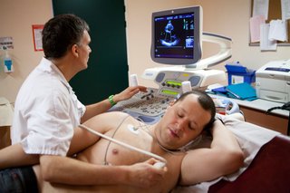
Before the scan, you need to remove the clothing on the top half of your body. You may be offered a hospital gown to cover yourself during the test. You lie down on your left side.
Your doctor will:
- attach several small sticky sensors (electrodes) to your chest to monitor your heart rhythm
- put a lubricating gel on your chest or the ultrasound probe
- move the probe across your chest
You may hear a swishing noise during the scan. This is the sound of the blood flow through your heart.
The procedure usually takes up to 25 minutes. You can go home soon after it is over.
Risks or side effects
A standard echocardiogram is a simple, painless, safe procedure. There are no side effects from the scan.
You may feel:
- cold from the lubricating gel
- some minor discomfort on your skin when the electrodes are removed
Other types of echocardiogram
Other types of echocardiogram include:
- transoesophageal echocardiogram (TOE)
- stress echocardiogram
- contrast echocardiogram
Transoesophageal echocardiogram (TOE)
Your doctor may request a TOE to get more detailed images. This can help to plan for heart surgery.
You may need to avoid eating for several hours before this test.
The doctor will:
- numb your throat with a local anaesthetic spray
- give you a sedative injection to help you relax
- pass a small probe down your throat into your food pipe and stomach
The doctor may also monitor your oxygen level during the test. This is to check for any breathing problems caused by sedation medicine.
Recovering after a TOE
You may find the TOE procedure uncomfortable.
You cannot eat or drink anything for at least 2 hours after the test, as the back of your throat will still be numb. When you have recovered you will be given a light snack.
Your throat may feel sore for a few hours afterwards. There's also a small chance of the probe damaging your throat.
If you have a sedative, you cannot drive, operate a machine, make important decisions or drink alcohol until the next day. You will need to arrange for someone to accompany you home. This is because you may still feel drowsy.
Stress echocardiogram
If your heart problem is triggered by physical activity, your doctor may do an echocardiogram:
- during or just after exercise on a treadmill or exercise bike
- after an injection of a medicine that makes your heart work harder
This is called a stress echocardiogram.
During a stress echocardiogram, you may:
- feel sick and dizzy
- have some chest pain
There's a small risk that the procedure triggers an irregular heartbeat or heart attack. But your doctor will monitor you during the test. They will stop the test if there are signs of any problems.
Contrast echocardiogram
This is where a harmless substance called a contrast agent is injected into your blood. This substance shows up clearly on the scan. It can help create a detailed image of your heart and see the blood flow better.
Some people have a reaction to the contrast agent. This often only causes mild symptoms such as itching.
Getting your results
You do not get your results straight away.
The specialist who does the scan usually:
- analyses the scan
- sends the results to the doctor who requested the test
Your doctor will discuss the results with you at your next appointment.
Content supplied by the NHS and adapted for Ireland by the HSE
